Clavicle Fracture
Clavicle Fracture
The shoulder is made up of three bones: the humerus (upper arm bone), the clavicle (collarbone), and the scapula (shoulder blade). The clavicle is a very commonly injured bone and can be injured during a fall onto the shoulder. There are several types of clavicle fractures, but the most common type occurs in the middle of the bone. Patients who have a clavicle fracture with feel pain at the top of the shoulder with movement of the arm and might feel clicking when moving the arm. Bruising or a bump can also be seen along the top of the shoulder.
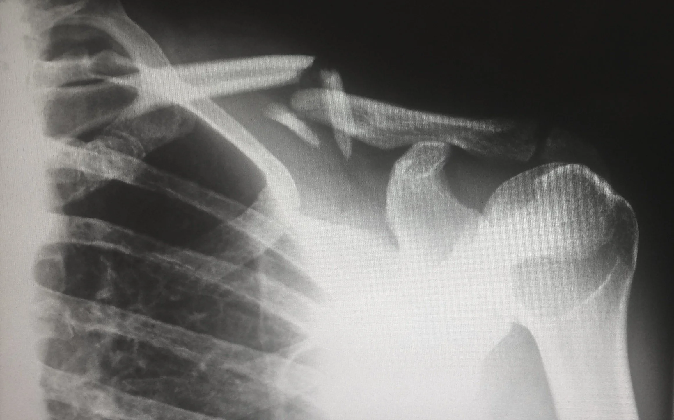
Clavicle fractures are diagnosed using physical examination and imaging. The physical examination focuses on identifying where the pain is located and examining the nerves and blood vessels of the arm. The skin over the top of the shoulder will also be examined for any skin tenting or for any areas where the bones pierced the skin. If any skin injury is found, surgery is required to protect the skin and decrease the risk of infection. X-rays will show the fracture and allow identification of the type and severity of the injury. The majority of clavicle fractures can be treated without surgery. Usually, MRI and CT are not needed for clavicle fractures.
Education
X-ray of the left shoulder showing a midshaft (middle) clavicle fracture.
The majority of clavicle fractures can be treated without surgery. A sling is used for the first 1-2 weeks for comfort. Patients will return for a repeat X-ray 2 weeks and 6 weeks after the injury to assess the location of the fracture pieces. Complete healing occurs between 8-10 weeks after the injury. Studies have shown that patients who smoke have slower bone healing and Dr. Kew encourages smoking cessation to help with healing and overall health. Physical therapy is used to strengthen the shoulder and patients are able to return to sports around 3 months after injury, if the shoulder has regained full strength and range of motion.
Surgery is used in severe fractures with significant overlap or displacement of the fragments. It can also be used in patients who where the clavicle healed in an unsatisfactory position (malunion) or did not heal (nonunion). The surgery performed is called an Open Reduction and Internal Fixation (ORIF) and is where a plate and screws is used to hold the fracture fragments in appropriate alignment. The plate and screws can stay forever, but about 30% of patients feel pain or sensitivity around the hardware. The plate and screws can be removed if symptoms develop after the fracture has healed.
Disclaimer: The information presented on this page has been prepared by Dr. Kew and should not be taken as direct medical advice, merely education material to enhance a patient’s understanding of specific medical conditions. Each patient’s diagnosis is unique to the patient and requires a detailed examination and a discussion with Dr. Kew about potential treatment options. If you have specific questions about symptoms you are having and would like to discuss with Dr. Kew, please click below to contact our office to schedule a visit. We hope the information above allows our patients to more thoroughly understand their diagnosis and expands the lines of communication between Dr. Kew and her patients.
