Anterior Cruciate Ligament Injury
ACL Injury
The knee is the most complex joint in your body and uses a delicately balanced system of bones, ligaments, and other structures to allow you to run, jump, perform cutting movements, and other advanced activities. The knee is made up of three bones: the femur (thigh bone), the tibia (shin bone), and the patella (kneecap). The two joints of the knee are the tibiofemoral joint (between the femur and tibia bones, this is the main knee joint) and the patellofemoral joint (between the kneecap and the trochlea, a groove on the front of the femur). The fibula bone is a thin bone on the outside of your lower leg that does not contribute to the knee joint, but is important to ankle stability.
Due to the geometry of the knee joint, it is inherently unstable and relies on ligaments, tendons, and other soft tissue (non-bony) structures for stability.
Ligaments: The knee ligaments support the knee through range of motion
Anterior cruciate ligament: This ligament is the most commonly discussed ligament, as it is commonly injured. It is located in the middle of the knee and runs from the back of the femur to the tibia. The ACL prevents the tibia from moving too far forward.
Posterior cruciate ligament: This ligament is also located in the middle of the knee and runs from the front of the femur to the back of the tibia. The PCL prevents the tibia from sliding too far backward.
Medial collateral ligament: This ligament is located along the inside of the knee and runs from a bony prominence on the inside of the femur to the inside of the tibia. It prevents the knee from collapsing inward.
Lateral collateral ligament: This ligament is located along the outside of the knee and runs from a bony prominence on the outside of the femur to the top of the fibula bone. This ligament works in concert with other soft tissue structures to prevent the knee from collapsing outward.
ACL injuries are very common and occur when the knee sustains a direct blow or, more commonly, during a non-contact injury when the knee rotates while the foot is planted. The ACL can be injured as a partial tear, or a portion of the ligament is torn, or a complete tear. ACL tears can occur in isolation, but are often seen with meniscus tears, cartilage injury, or injury to other ligaments.
Education
Risk Factors
ACL injuries are very common in the athletic population, but specific risk factors have been found that can increase a patient’s risk of injury.
Female athletes are 4 times more likely to sustain an ACL injury than male athletes. This is due to differences in anatomy, neuromuscular forces, ratio of quadriceps and hamstring muscle strength, and other factors currently under investigation.
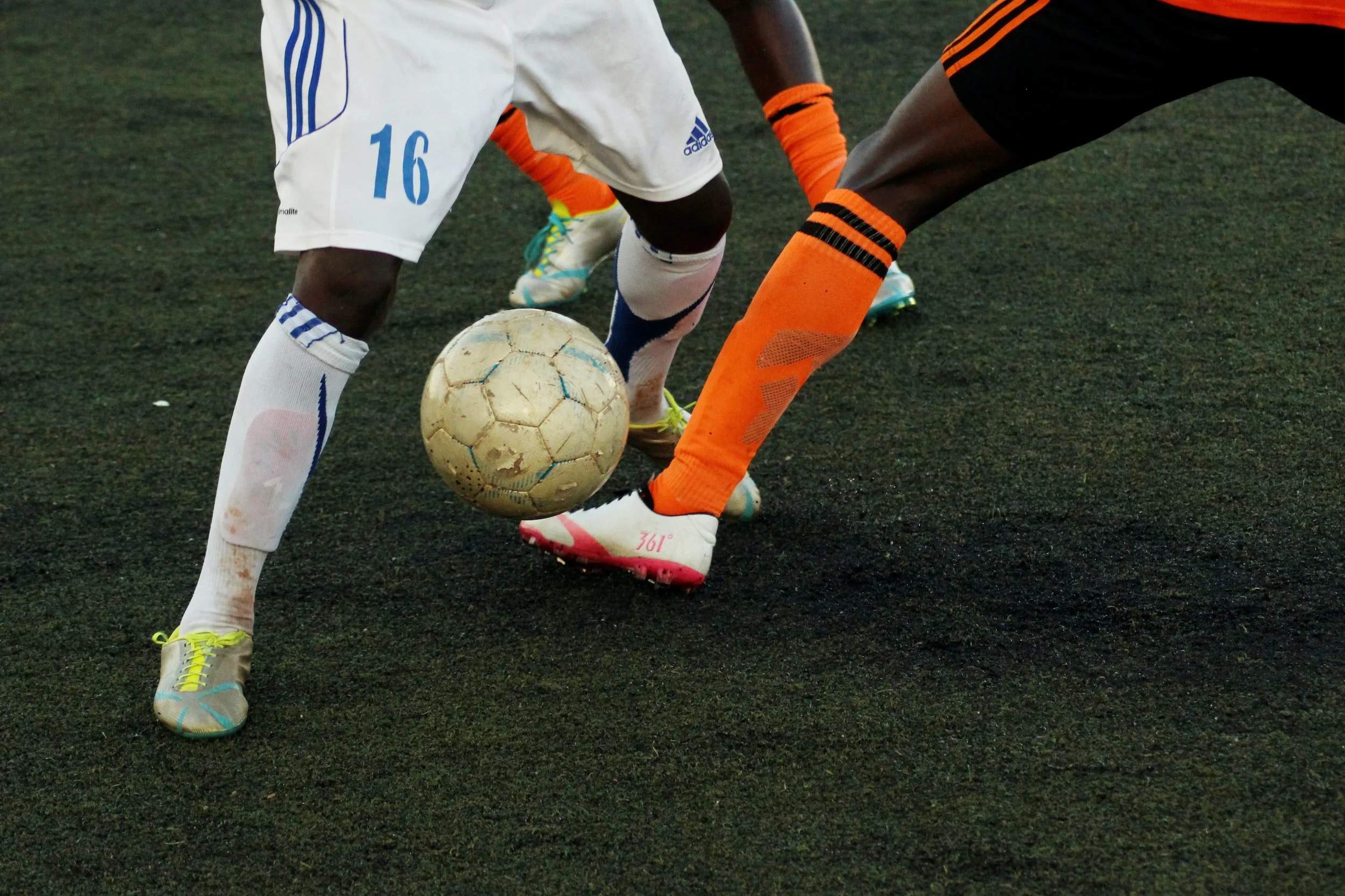
Patients who play a sport that involves side to side cutting movements have an increased risk of sustaining an ACL tear. Soccer players have been found to be at a uniquely high risk of an ACL injury.
Patients who have a history of an ACL injury also have an increased risk of sustaining a second ACL injury, either to the previously injured knee or to the other knee
Symptoms
Patients may feel a pop in the knee during an ACL injury and will not be able to continue participating in activity. During the first 12-24 hours, patients will notice knee swelling and difficulty walking due to the swelling. Occasionally, patients may notice bruising around the knee. After the knee swelling subsides, patients will notice minimal symptoms to the knee. They will be able to walk normally and will not have much, if any, pain.
Diagnosis
ACL injuries are diagnosed using physical examination and imaging studies to confirm the diagnosis. The physical examination is used to identify areas of pain, swelling, change in range of motion, and laxity (or looseness) of the knee. X-rays are used to look at the bones of the knee. MRI (magnetic resonance imaging) is an important part of the diagnosis and will allow evaluation of all the knee ligaments, meniscus, cartilage, and any other structures that may have been injured.
Treatment
There is a common misconception that all ACL injuries require surgery to allow the patient to return to full activities. Treatment of an ACL injury should be individualized to each patient based on age, symptoms, associated injuries, and activity goals. Non-surgical treatment of ACL injuries can be used in patients who are active, but do not participate in cutting sports, or those who lead a less active lifestyle. Non-surgical treatment of ACL injuries can include:
Rest and activity modification: All sports and active pursuits should be stopped until the knee inflammation resolves. Occasionally, patients require several weeks of crutch use to regain a normal gait.
Non-steroidal anti-inflammatory medications (NSAIDs) can help decrease inflammation
Brace: Use of a knee brace can help the patient feel more confident and secure when walking
Physical therapy: This is an important tool to help you strengthen and retrain the muscles of the knee to work better together
Surgical treatment is recommended for young, active patients and those who participate in cutting or pivoting sports. Patients with an ACL tear who will undergo surgery will complete a course of physical therapy before surgery to work on decreasing inflammation, regaining normal knee range of motion, and leg strengthening exercises. ACL surgery usually occurs several weeks after the injury to ensure that normal knee range of motion is obtained. ACL reconstruction has been shown to be the most reliable surgery to allow a return to full activities with a stable and functional knee.
ACL reconstruction is performed using arthroscopy and you will have small incisions that are used for the camera and specialized instruments
Reconstruction of the ACL can be done with several different graft options which are discussed below
Patients who receive an autograft (their own tissue) will also have an incision from where the graft is taken
The graft is placed into tunnels that are drilled in the tibia and femur bones and is secured with screws or a metal button to hold the graft in place
Patients are able to walk after the procedure on crutches (unless other surgeries are performed at the same time on the meniscus, cartilage, etc).
Rehabilitation is a critical part of the recovery from an ACL injury and will focus on regaining quadriceps strength and symmetry to the uninjured leg
Patients will undergo testing during their recovery to ensure that specific strength, range of motion, and functional metrics are being met
Patients are able to return to full activities between 9-12 months after their surgery, depending on the results of the testing and a discussion with Dr. Kew
ACL Graft Options
The ACL can be reconstructed using several different graft types and we’ll discuss each one below. Autograft tissue is taken from the patient’s own knee, while allograft tissue is from a deceased donor. Graft selection is based on many factors, including patient age, activity level, and other factors.
Bone-patellar tendon-bone autograft (BTB)
This graft is taken from the patient’s own knee and is harvested from the middle third of the patellar tendon (runs under the kneecap to the tibia) and includes bone pieces on each end that are taken from the bottom of the kneecap and top of the tibia
The BTB graft allows for bone to bone healing of the graft over time
This graft is the most commonly used graft in elite athletes and has the longest successful track record in orthopaedic literature
Some disadvantages to this graft include:
Knee pain with kneeling or patellar tendinitis after surgery can develop in some patients
Patients may experience higher levels of pain immediately after surgery due to the bone pieces that were taken from the patella and tibia
Larger incision is needed to harvest the graft
There is a small risk of a patella fracture after surgery. This is a rare occurrence, but is of great concern to surgeons
Hamstring tendon autograft
This graft is taken from the patient’s hamstring tendons, which are located on the inside of the lower leg
Taking these hamstring tendons does not affect your ability to walk or participate in any activities or sports
Mild hamstring weakness has been noted in patients who are sprinters or those who require quick acceleration in their sport
Generally, 2 of the 3 available tendons are taken and weaved together to create a new ACL graft
Patients will have less pain than those who have a BTB graft and may be able to return to walking with a normal gait faster
The most important disadvantage to the hamstring graft is weaker initial fixation. The graft heals well over time, but initial physical therapy and activity should be carefully monitored to protect the graft
Quadriceps tendon autograft
This graft is taken from the patient’s quadriceps tendon (runs from the thigh muscles to the top of the patella) and the central 1/3 of the tendon is taken
This graft can be taken with a bone piece from the patella or just as a soft tissue graft (no bone)
Quadriceps tendon autograft is the newest graft in the literature, but has been shown to be equivalent to the other autograft options. Due to its recent use, we don’t yet have long-term outcomes (>10-20 years) of patients who receive a quadriceps tendon autograft
Patients will have a similar postoperative pain course as patients who receive a hamstring graft
Quadriceps tendon autograft has a similar disadvantage as hamstring tendon autografts, as initial fixation is not as robust and early physical therapy will be monitored to protect the graft
Allograft
This graft is taken from a deceased donor and several different tendon options can be used
Patients will only have the incisions required for the ACL reconstruction, as there is no graft harvest incision
Patients are able to return to walking normal and daily activities quicker due to the decreased pain and swelling after surgery
Disadvantages of using allograft tissue include:
Viral infection from the allograft tissue can occur, but is exceedingly rare and tissues are carefully sterilized and examined to ensure no viral transmission will occur
Studies have shown a significantly higher rate of ACL graft failure in young patients who participate in cutting and pivoting sports
Disclaimer: The information presented on this page has been prepared by Dr. Kew and should not be taken as direct medical advice, merely education material to enhance a patient’s understanding of specific medical conditions. Each patient’s diagnosis is unique to the patient and requires a detailed examination and a discussion with Dr. Kew about potential treatment options. If you have specific questions about symptoms you are having and would like to discuss with Dr. Kew, please click below to contact our office to schedule a visit. We hope the information above allows our patients to more thoroughly understand their diagnosis and expands the lines of communication between Dr. Kew and her patients.

