Shoulder Dislocation/ Instability
Shoulder Instability
The shoulder is one of the most complex joints in your body and allows your arm to rotate and move in many different directions. The shoulder joint is described as a “ball and socket” joint, however, the socket is very shallow and is similar to a golf ball resting on a tee. The shoulder is made up of three bones: the humerus (upper arm bone), the clavicle (collarbone), and the scapula (shoulder blade). The outer portion of the scapula bone is called the glenoid and is the golf tee where the ball of the humeral bone rests to make up the shoulder joint.
Due to the geometry of the shoulder joint, it is very flexible and allows a large range of motion, but this leads to an inherent risk of injury to the shoulder. Several soft tissue structures (non-bony structures) exist in the shoulder joint to improve the stability of the joint.
Labrum: A rim of cartilage on the shoulder socket (glenoid) that helps stabilize the joint by providing a bumper for the ball around the socket.
The labrum can be damaged when a shoulder dislocates, creating a Bankart lesion, or injury to the front of the labrum
A Hill-Sachs lesion can also occur with a shoulder dislocation and refers to the bony damage to the back of the humeral head from contact with the glenoid during the dislocation
Capsule: Every joint has an envelope around it to contain the joint and the joint fluid.
Ligaments: Several ligaments in the shoulder joint strengthen the capsule and increase stability of the shoulder joint
Rotator cuff muscles: A group of 4 muscles that are an important contributor to shoulder stability and function. Each muscle attaches to the humeral head (ball of the shoulder joint) and work in a coordination fashion to keep the ball centered in the socket
The subscapularis is a large rotator cuff muscle that attaches to the front of the ball and allows you to rotate your arm inward
The supraspinatus and infraspinatus muscles attach at the top of the ball and allow you to raise your arm overhead
The teres minor attaches to the back of the ball and allows you to rotate your arm outward
Other soft tissue structures in the shoulder include:
Bursa: These structures are located between bone and the surrounding soft tissue above the rotator cuff. They serve as a cushion between the rotator cuff and the bones of the shoulder
Cartilage: This structure is present in all joints, where its called articular cartilage, and covers both ends of the bones in the joint. Cartilage allows the bones to glide easily as the joint is moved.
The axillary nerve is a large nerve that provides motor and sensation to the outside of the shoulder. This nerve can be stretched or damaged during a shoulder dislocation. The injury to the nerve is typically temporary and will be closely monitored during your visits. Timing of surgery, if needed, may change if the axillary nerve is injured
Types of Instability
Shoulder instability refers to a condition where the shoulder comes out of the socket easily or if there is a sense of looseness in the shoulder with specific arm positions. Instability can occur from a traumatic event, such as a fall, or can be due to a biologic looseness to the shoulder.
The shoulder can dislocate to the front (anterior instability), to the back (posterior instability), or can be multi-directional. There are different degrees of dislocation and the shoulder can partially dislocate (subluxation) or fully dislocate. Anterior shoulder instability is the most common type.
Dislocation usually occurs during a forceful twisting outward motion (external rotation) when the arm is above the level of the shoulder. Some dislocated shoulders will pop back in on their own, while others may require a “reduction”, which is when another person puts it back in place. Age is the most important risk factor for recurrent shoulder instability (another dislocation or subluxation). The rate of recurrent instability without surgery is more than 90% in those younger than 20 years, 60% for those between 20 and 40 years, and less than 10% if over 40 years old. Patients over the age of 40 who sustain a shoulder dislocation have an increased risk of developing a rotator cuff tear during the dislocation. 30% of patients over the age of 40 years, and 80% of patients over the age of 60 years have been found to have a rotator cuff tear after a shoulder dislocation.
Education
Risk Factors for Shoulder Instability
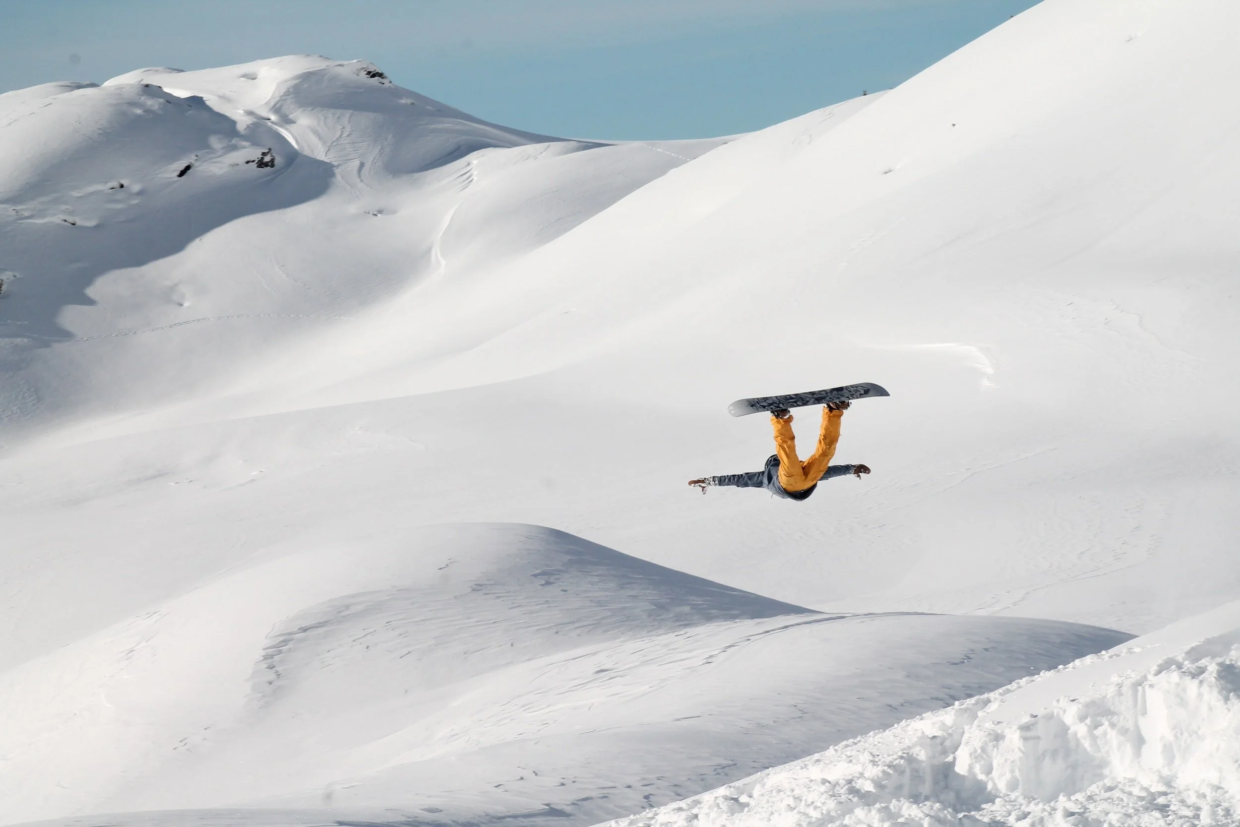
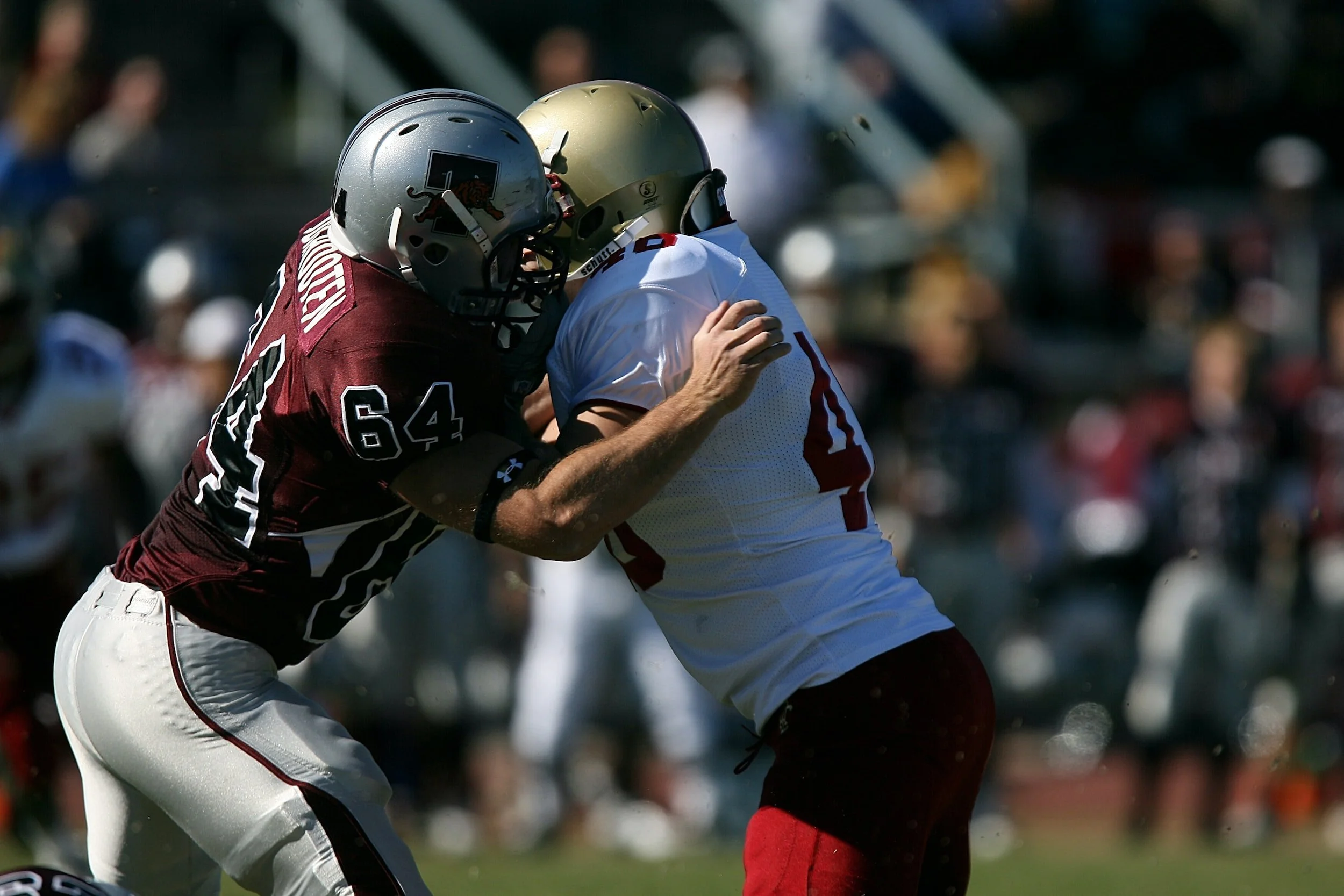
Patients who play contact sports or participate in high level activities, such as snowboarding, skiing, mountain biking, are at risk for shoulder dislocations. Typically, anterior shoulder dislocations occur in this population of patients.
Specific populations of patients have a higher risk of developing posterior shoulder instability due to repetitive microtrauma, such as football linemen or patients who are very active in weight lifting. Patients with a seizure disorder are at risk for shoulder instability due to the traumatic nature of a seizure and present a unique treatment challenge to physicians.
Patients with a history of ligamentous laxity, or increased flexibility, can also develop shoulder instability due to the inherent looseness of their joints. This type of instability is called multi-directional instability (MDI) and responds well to non-surgical treatment to strengthen the muscles that control the shoulder and shoulder blade. This condition can require a long rehabilitation process, as it takes time to retrain the muscles of the shoulder .
Symptoms
At the time of injury, patients may not be able to move the shoulder and may see a prominence of the front of the shoulder. Patients with shoulder instability can feel a sense of looseness of the shoulder, especially in specific arm positions (when mimicking a pitching motion). There may also be apprehension, or fear, to do certain activities because of the loose feeling in the shoulder.
Diagnosis
Shoulder instability is diagnosed using physical examination and imaging studies to confirm the diagnosis. The physical examination is used to identify areas of pain, weakness, and identify if certain positions cause apprehension. The integrity of the axillary nerve will also be examined.. X-rays help determine if the shoulder is reduced (in place) and if there are any bony injuries. MRI (magnetic resonance imaging) allows us to evaluate the soft tissue and non-bony parts of your shoulder and specifically look at the labrum, biceps tendon, and rotator cuff. Occasionally, a CT (computed tomography scan) will be needed to look at the bony architecture of the shoulder. This is needed if a patient has had multiple dislocations to the same shoulder or if Dr. Kew needs to closely evaluate the bones of the shoulder.
Treatment
Treatment options for shoulder instability are tailored to the individual patient and take into account the patient’s age, desired activity level, and history of shoulder dislocation, among other factors. General descriptions of a non-surgical and surgical treatment plan are detailed below.
Nonoperative
A non-surgical approach is reasonable in many patients, especially after a first-time dislocation. The shoulder is placed in a sling to provide support and immobilization for 1-2 weeks and physical therapy is started to work on shoulder strengthening and range of motion. Any sports or athletic activity are restricted until the shoulder has regained range of motion and strength. You will be cleared by Dr. Kew to ensure that the shoulder has recovered and is strong enough to return to activity.
Surgical
Surgery is often needed in younger patients who have a multiple shoulder dislocations or have tried non-surgical treatment and occasionally, in patients with a first-time dislocation. Surgical options include:
Arthroscopic labral repair: This procedure is performed in patients who have a tear in the front or back of the labrum after a dislocation. Small incisions are made in the shoulder, through which a camera and specialized instruments are used to repair the labrum and capsule back to the glenoid using small anchor devices and stitches.
Open bony reconstruction: Patients who have multiple shoulder dislocations may wear out the bone of the shoulder (glenoid). If this occurs, reconstruction of the lost bone can be done with a procedure called a Latarjet. This procedure takes a small piece of bone from the front of the shoulder and places it next to the glenoid to create a new shoulder joint. Other procedures may be required if the size of the missing bone is very large. If these procedures are required for your shoulder instability, Dr. Kew will go through them in detail with you during your visit.
Disclaimer: The information presented on this page has been prepared by Dr. Kew and should not be taken as direct medical advice, merely education material to enhance a patient’s understanding of specific medical conditions. Each patient’s diagnosis is unique to the patient and requires a detailed examination and a discussion with Dr. Kew about potential treatment options. If you have specific questions about symptoms you are having and would like to discuss with Dr. Kew, please click below to contact our office to schedule a visit. We hope the information above allows our patients to more thoroughly understand their diagnosis and expands the lines of communication between Dr. Kew and her patients.

